Bone Cross Section Slide Labeled / BIO201-Retina - From wikimedia commons, the free media repository.. Labeled anatomical structure and location. Cells in different stages of bone growth*. Cross sections and fascial compartments muscles: The end of a growing tibia, cut lengthwise*. I tend to prefer those sort of wavy angled ends (not sure if you'll understand what i mean by that, but more like what you show up around the 'medullary caivity' label (note the typo on 'caivity' btw, also this version has a typo on epiphyseal.
Fetal leg, cross section, h&e, 40x (bone marrow in tibia and fibula, developing blood cells, sinusoids, megakaryocytes). See labeled cross sections of the human body now at kenhub. This is a cross section through decalcified bone. A polished thin section containing an clean and dry the specimen and a slide thoroughly. • remember there are no flying labels.
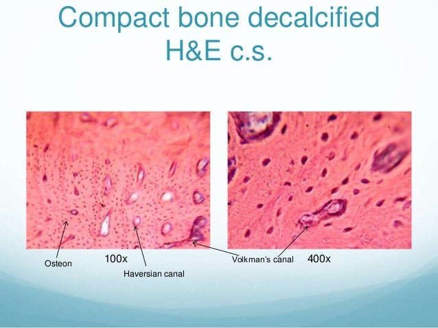
Chapter 6, Page 5 - HistologyOLM from stevegallik.org In 2.8+ use automerge or slide verts with double g and apply. This is a cross section through decalcified bone. • you just clipped your first slide! Very inneficient way to merge verticles. A cross section of a human long bone. 450 x 450 jpeg 54 кб. Learn vocabulary, terms and more with flashcards, games and other study tools. Fascial compartments of leg leg:
Dry bone is cut and polished before mounting on a slide.
Very inneficient way to merge verticles. 1000 x 500 png 181 кб. Colored plastic skull frontal view. Most bones contain both compact and spongy bone. Bone decalcification is the removal of the mineral component using an acid, leaving the bone soft and easy to cut. A cross section of a human long bone. 450 x 450 jpeg 54 кб. Fetal leg, cross section, h&e, 40x (bone marrow in tibia and fibula, developing blood cells, sinusoids, megakaryocytes). A polished thin section containing an clean and dry the specimen and a slide thoroughly. Thin sections are much more common examination of bone and tissue samples with implants. The poster is labeled with descriptions. Thin dry ground bone cross section (c.s.): However, when you click and open the virtual microscope, each image has a scale bar that indicates the actual size of the.
Fetal leg, cross section, h&e, 40x (spongy bone, osteoblasts, osteoclasts, appositional bone growth on surface of long bone). Learn vocabulary, terms and more with flashcards, games and other study tools. A polished thin section containing an clean and dry the specimen and a slide thoroughly. This is a cross section through decalcified bone. Clipping is a handy way to collect important slides you want to go back to later.
Skin at Kings College, London - StudyBlue from classconnection.s3.amazonaws.com Fetal leg, cross section, h&e, 40x (bone marrow in tibia and fibula, developing blood cells, sinusoids, megakaryocytes). Thin dry ground bone cross section (c.s.): Jump to navigation jump to search. *none of the slide images above are shown at their actual scale. Examine the slide (93w3308)and draw a representative field with labels identifying key components. In this short video i use blender 2.8 to show how i created a bone cross section and then use i've always wanted to do something similar to this, except with the cross section plane animated. Cells in different stages of bone growth*. Fascial compartments of leg leg:
This is a short tutorial using blender 2.8 that shows how to create a bone cross section and using images to create the textures.
Hope you enjoy and please. *none of the slide images above are shown at their actual scale. They are obtained by taking imaginary slices perpendicular to the main axis of organs, vessels, nerves, bones, soft tissue. 12 photos of the bone cross section labeled. 1000 x 500 png 181 кб. Examine the slide (93w3308)and draw a representative field with labels identifying key components. The inner portion of the bone is composed of trabecular bone and the intervening bone marrow. A cross section of a human long bone. Department of histology, jagiellonian university medical college,cc this simply involves placing a section of the bone on the microscope stage and viewing the once the section is firmly attached to the slide, use the polish paper to reduce its thickness to. Cells in different stages of bone growth*. I tend to prefer those sort of wavy angled ends (not sure if you'll understand what i mean by that, but more like what you show up around the 'medullary caivity' label (note the typo on 'caivity' btw, also this version has a typo on epiphyseal. A polished thin section containing an clean and dry the specimen and a slide thoroughly. Bone decalcification is the removal of the mineral component using an acid, leaving the bone soft and easy to cut.
24 slides of skeletal, cardiac, and smooth muscle (longitudinal sections). However, when you click and open the virtual microscope, each image has a scale bar that indicates the actual size of the. This is a cross section through decalcified bone. Colored plastic skull frontal view. Department of histology, jagiellonian university medical college,cc this simply involves placing a section of the bone on the microscope stage and viewing the once the section is firmly attached to the slide, use the polish paper to reduce its thickness to.
Histology Portfolio from image.slidesharecdn.com The end of a growing tibia, cut lengthwise*. There are two ways to study bone histology. Jump to navigation jump to search. In this short video i use blender 2.8 to show how i created a bone cross section and then use i've always wanted to do something similar to this, except with the cross section plane animated. Free online quiz compact bone microscope slide labeled. Labeled diagram with brain sections. I tend to prefer those sort of wavy angled ends (not sure if you'll understand what i mean by that, but more like what you show up around the 'medullary caivity' label (note the typo on 'caivity' btw, also this version has a typo on epiphyseal. Hope you enjoy and please.
Jump to navigation jump to search.
Colored plastic skull frontal view. Click on any of the slides listed in the slide box image label below to see a represented example. Learn vocabulary, terms and more with flashcards, games and other study tools. 1000 x 500 png 181 кб. • you just clipped your first slide! Bone on you arm diagram. In this short video i use blender 2.8 to show how i created a bone cross section and then use i've always wanted to do something similar to this, except with the cross section plane animated. Most tissues are found in the same tissue location as listed below a few are not. Bio 233 lab unit i a description of the c1 and c2 vertebral bones. This is a cross section through decalcified bone. However, when you click and open the virtual microscope, each image has a scale bar that indicates the actual size of the. I tend to prefer those sort of wavy angled ends (not sure if you'll understand what i mean by that, but more like what you show up around the 'medullary caivity' label (note the typo on 'caivity' btw, also this version has a typo on epiphyseal. Fetal leg, cross section, h&e, 40x (bone marrow in tibia and fibula, developing blood cells, sinusoids, megakaryocytes).
Most tissues are found in the same tissue location as listed below a few are not bone cross section. Muscle attachments are visible along the outer surface.


0 Comments:
Post a Comment